Background introduction Acute promyelocytic leukemia (APL) is a specific subtype of acute myeloid leukemia. In cytogenetics and molecular biology, APL has a characteristic t(15;17)(q22;21) translocation, forming a PML-RARA fusion gene. A large number of data indicate that patients carrying the PML-RARA fusion gene are predictive of sensitivity to ATRA therapy and good clinical efficacy.
Dwarka Sector-7, New Delhi-110075
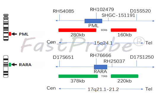
PML/RARA gene fusion probe detection kit
Probe description
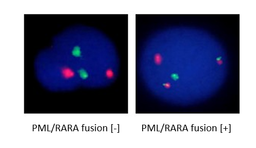
The two probes bind to the target detection site by in situ hybridization using an orange-red fluorescein-labeled PML probe and a green fluorescein-labeled RARA probe. Under normal conditions (the PML/RARA gene is not fused), it shows two orange-red signals and two green signals under a fluorescence microscope. When a fusion gene is present, the green and orange-red signals form a yellow fusion signal due to recombination.
Clinical significance
PML/RARA gene fusion is a hallmark of acute promyelocytic leukemia (APL). PML/RARA protein fusion inhibits the differentiation and maturation of promyelocytic cells by dominant negative inhibition, thereby blocking cell differentiation leading to sustained proliferation. All-trans retinoic acid (ATRA) and arsenic trioxide can target the degradation of PML/RARA fusion protein, restore the function of wild-type PML and RARA genes, relieve their inhibition of gene transcription, induce cell differentiation and apoptosis, and effectively treat APL. The combination of ATRA and chemotherapy can achieve a complete response rate of 90% to 95% of APL, and can achieve long-term survival of more than 70% of patients.

AML1/ETO gene fusion probe detection kit
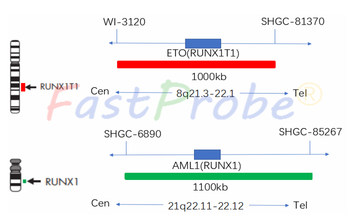
Background introduction AML1/ETO gene fusion formed by chromosome 8 and chromosome 21 translocation is a common cytogenetic abnormality in patients with acute myeloid leukemia (AML), and about 12% to 20% of patients with acute myeloid leukemia have AML1/ETO gene fusion. While the positive rate of AML-M2 leukemia is 20% to 40%, and the positive rate of M2b subtype is as high as 90%, which is rare in other types of leukemia. The AML1/ETO protein fusion is a transcriptional repressor that inhibits normal AML1 protein-mediated function, alters the process of self-renewal and maturation of hematopoietic progenitor cells, and also signals the initiation of abnormal hematopoietic cell proliferation, causing the proliferation of leukemia cells
Probe description
ETO probe is labeled with an orange-red fluorescein, and AML1 probe with a green fluorescein. The two probes combine to the target detection site by in situ hybridization. Under normal conditions (AML1/ETO gene is not fused), it shows two orange-red signals and two green signals under a fluorescence microscope. When a fusion gene is present, the green and orange-red signals form a yellow fusion signal due to recombination.
Clinical significance
AML1/ETO gene fusion can be used as AML diagnostic assistant and prognosis assessment means. Clinically, t(8;21) leukemia represents a type of acute leukemia with good prognosis. Adult patients have good response to treatment, high complete remission rate, long median survival time, but prone to recurrence. Children’s treatment and prognosis are not as good as adult patients.
MLL gene break apart probe detection kit
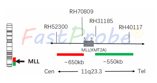
Background introduction The MLL (Mixed-linage leukemia or Myeloid-lymphoid leukemia) gene is located in the No. 11 staining map, Zone 2, Zone 3 (11q23), and was successfully cloned as early as 1991. The MLL gene is a key gene in the regulation of hematopoietic processes, and its abnormality is closely related to the pathogenesis of leukemia. According to statistics, there are at least 104 MLL gene rearrangements, and up to 64 MLL genes fusion have been identified. Most of the leukemia’s with MLL gene fusion are highly malignant, not sensitive to chemotherapy, and have low remission rate. Therefore, the detection of MLL gene fusion in acute leukemia is of great significance for the choice of treatment options for leukemia, residual lesion detection and prognosis.
Probe description
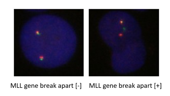
MLL gene region 5’ end is labeled with an orange-red fluorescein, and the 3' end of MLL gene labeled with a green fluorescein. MLL gene break apart probe is used to detect 11q23 segment translocation, and all MLL gene rearrangements could be detected, avoiding separate detection or missed diagnosis caused by gene fusion.
Clinical significance
Common translocation forms of the MLL gene are t(4;11), t(9;11), t(11;19) and other recombination’s. 8-10% of acute myeloid leukemia (AML) has this abnormality. MLL recombination exists in 80% of infants with AML, suggesting a moderate risk type; other MLL genes are recombined into high-risk types.

CBFB/MYH11 gene fusion probe detection kit
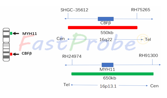
Background introduction Acute myeloid leukemia (AML) is a group of highly heterogeneous hematopoietic malignancies, often associated with acquired chromosomal abnormalities, the most common of which is chromosomal translocation. Chromosomal inversion of inv16 (p13q22) or translocation t(16;16) (p13; q22) found in myeloid leukemia (AML-M4) cells with eosinophilia, resulting in the MYH11 gene located at 16p13. The CBFB gene located at 16q22 is recombined to form a CBFB/MYH11 gene fusion. The detection rate of CBFB/MYH11 gene fusion in myeloid leukemia is about 7%. Since the CBFB/MYH11 gene fusion is only found in AML, according to the WHO leukemia diagnostic criteria, AML can be diagnosed by detecting the CBFB/MYH11 gene fusion.
Probe description
CBFB probe is labeled with an orange-red fluorescein, and MYH11 probe is labeled with a green fluorescein. The two probes combine to the target detection site by in situ hybridization. Under normal conditions (CBFB/MYH11 gene did not fuse), it shows two orange-red signals and two green signals under a fluorescence microscope. When a fusion gene is present, the green and orange-red signals form a yellow fusion signal due to recombination. The method was used to detect the status of CBFB/MYH11 gene fusion providing a reference for the identification, prognosis and drug administration guidance for clinical AML leukemia patients.
Clinical significance
CBFB/MYH11 gene fusion can be used for the diagnosis of AML. In addition, in the case of positive CBFB/MYH11 gene fusion, the detection of CBFB/MYH11 gene fusion has become the most valuable indicator for the determination of therapeutic options and therapeutic efficacy evaluation. For example, quantitative analysis of CBFB/MYH11 gene fusion can also be used to judge the level of leukemia cells in patients, the detection of minimal residual disease and the prediction of recurrence risk. AML patients with CBFB/MYH11 gene fusion have a better prognosis, and high DFS and low recurrence rates can be achieved by HDAC regimen
CBFB gene break apart probe detection kit
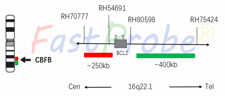
Background introduction CBFB gene break apart is a characteristic chromosomal abnormality of AML, accounting for 5%-10% of total AML patients and 23% of M4 patients It is usually found in the AML-M4E0 subtype, but less in M2, M5 and M4 (no eosinophilic granulocytosis). It is now considered that CBFB gene break apart is a characteristic genetic alteration of M4E0.
Probe description
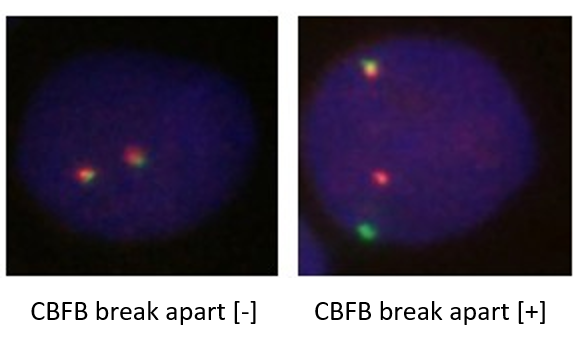
CBFB gene 5'end region uses an orange-red fluorescein, the CBFB gene 3'end uses a green fluorescein, and the translocation of 16q22 region is detected with MLL gene break probe. All CBFB gene rearrangements can be detected and avoiding separate detection due to missed diagnosis caused by gene fusion.
Clinical significance
Most AML patients with CBFB gene break apart are sensitive to chemotherapy and have a good prognosis