Background introduction TEL/AML1 dual-color double fusion probe aims to detect the translocation of the ETV6 (TEL) gene in chromosome 12p13.2 region and the RUNX1 (AML1) gene in the region of chromosome 21q22.12. The t(12;21)(p13.2;q22.1) translocation leads to the fusion of ETV6/RUNX1, the most common genetic recombination in patients with acute lymphoblastic leukemia (ALL) and is associated with a good prognosis. It is the highest incidence of childhood leukemia. In pediatric leukemia, acute lymphoblastic leukemia accounts for about 75%. In children with acute lymphoblastic leukemia aged 2-10, the positive rate of ETV6/RUNX1 (TEL/AML1) gene fusion accounts for about 20-25%, among which female is higher than male
Dwarka Sector-7, New Delhi-110075
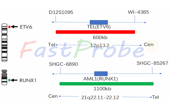
TEL/AML1 (ETV6/RUNX1) gene fusion probe detection kit
Probe description
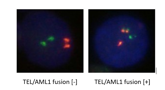
TEL probe uses an orange-red fluorescein label, and AML1 probe uses a green fluorescein label. The two probes combine to the target detection site by in situ hybridization. Under normal conditions (TEL/AML1 gene is not fused), it shows two orange-red signals and two green signals under a fluorescence microscope. When there is fusion, the green and orange-red signals form a yellow fusion signal due to recombination.
Clinical significance
TEL/AML1 gene fusion has a 20-25% incidence in children with B-ALL. It has a good prognosis, but is prone to recurrence.


MYC break apart gene probe detection kit
Background introduction MYC proto-oncogene is located on chromosome 8q24 and encodes a transcription factor that regulates cell growth. It is mainly activated by amplification and chromosomal translocation, and its downstream target genes affect cell proliferation, DNA and protein synthesis and metabolism.
Probe description
MYC dual-color break apart probe is a two directly labeled hybrid probe that hybridizes at the 8q24.21 region. The probe is directly labeled with an orange-red fluorescent dye that hybridizes with the proximal end of the MYC gene, and with a green fluorescent dye that hybridizes with the distal end of the MYC gene.
Clinical significance
Abnormal MYC gene break apart occurs in 5% of B-ALL patients and can fuse with multiple genes. Approximately 75% of mature B-cell acute lymphocytic patients are morphologically characterized by ALL-L3, often accompanied by a typical t(8;14) (q24; q32). Abnormal MYC gene break apart means that the prognosis is extremely poor and clear in clinical practice.
MLL break apart gene probe detection kit
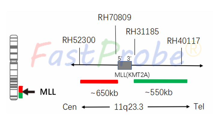
Background introduction MLL (Mixed-linage leukemia or Myeloid-lymphoid leukemia) gene located at 11q23 was successfully cloned as early as 1991. The MLL gene is a key gene in the regulation of hematopoietic processes, and its abnormality is closely related to the pathogenesis of leukemia.
Probe description
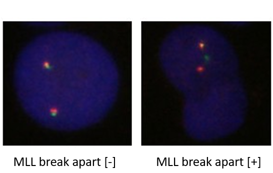
MLL gene 5'end region is labeled with an orange-red fluorescein and the 3'end labeled with a green fluorescein. The translocation of 11q23 region is detected with MLL gene break probe. All MLL gene rearrangements can be detected and avoiding separate detection due to missed diagnosis caused by gene fusion.
Clinical significance
MLL gene can fuse with 51 genes after chromosomal translocation. The incidence of MLL gene changes in acute leukemia is about 5%-10%, but in infant ALL it is up to 79%, which is a sign of poor prognosis. EFS in 5 years is only 26.7%.
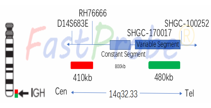
IGH break apart gene probe detection kit
Background introduction IGH separated dual-color probe aims to detect the translocation of 14q32.33 chromosome region (i.e., the IGH gene). IGH gene rearrangement is found in about 50% of NHLs (non-Hodgkin's lymphoma), and also in T-ALL, CLL and ALL. Studies have shown that IGH gene translocation also occurs in children's T-ALL.
Probe description
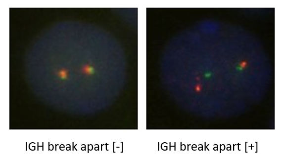
5'end of IGH gene region is labeled with an orange-red fluorescein, and the 3'end labeled with a green fluorescein. The translocation of 14q32 region is detected with IGH gene break probe. All IGH gene rearrangements can be detected and avoiding separate detection due to missed diagnosis caused by gene fusion.
Clinical significance
In ALL, the ratio of IGH to C-MYC translocation is the highest. In B-ALL and T-ALL, translocation of IGH with other genes is also more common.
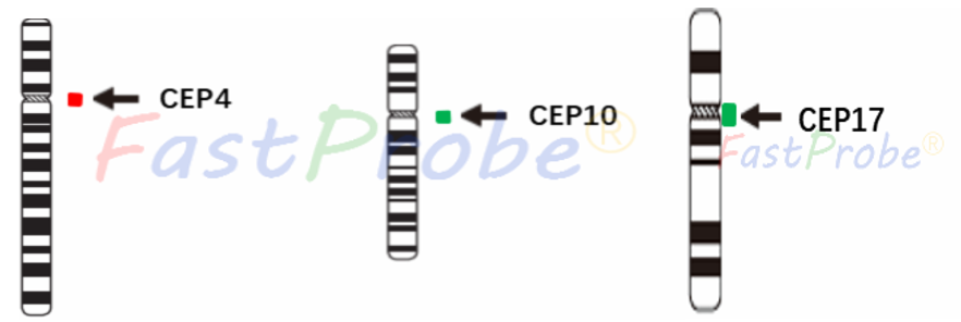
Chromosomes 4, 10 and 17 probe detection kit
Background introduction About 25% of children with ALL have an increase in the number of chromosomes, and chromosomes 4, 5, 6, 10, 17 and 21 are more common, among which trisomy 10 is the most common.
Probe description
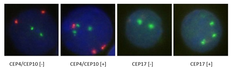
Chromosome 4-centromere region is labeled with an orange-red fluorescein, and the centromere region of chromosomes 10 and 17 is labeled with a green fluorescein. In normal cells, two orange-red signals and two green signals are observed under fluorescence microscopy. When chromosome number abnormality exists, three orange-red signals or three green signals are observed.
Clinical significance
The 4, 10, and 17 trisomy are independent prognostic indicators, and these patients have a 7-years EFS greater than 90%. The method of detecting the number of CEP4/CEP10/CEP17 chromosomes provides a reference for the clinical identification, prognosis and medication of leukemia patients.
P16 gene deletion probe detection kit
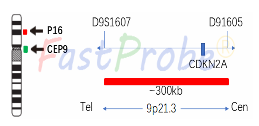
Background introduction P16 gene is located on the 9p21 chromosome and is a tumor suppressor gene. P16 gene deletion is present in 10% of ALL patients and has a higher proportion in T-ALL. Currently, FISH technology is widely used in the diagnosis of P6 gene deletion in ALL.
Probe description
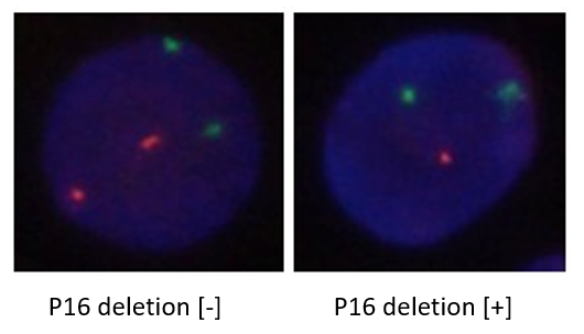
P16 gene deletion probe uses an orange-red dye to label P16 gene region, and a green dye is to label chromosome 9 centromere region (CEP9).
Clinical significance
One of the most common abnormalities in ALL is that homozygous deletions are mostly in T-ALL, and the proportion of homozygotes and heterozygotes in B-ALL is comparable; the prognosis is poor.