Background introduction BCL2 is a tumor suppressor gene located in the 18q21 region. BCL2 gene encodes a mitochondrial membrane protein that regulates apoptosis and is expressed in B cells. Translocation of the BCL2 gene is usually recognized in B cell lymphoma. In particular, translocation of t(14;18)(q32.3;q21.3) is present in approximately 80% of follicular lymphoma (FM), 20%-30% of diffuse large B-cell lymphoma In (DLBCL), it rarely occurs in B-cell chronic lymphocytic leukemia (B-CLL). Therefore, the detection of BCL2 translocation by fluorescence in situ hybridization (FISH) may have diagnostic and prognostic significance.
Dwarka Sector-7, New Delhi-110075
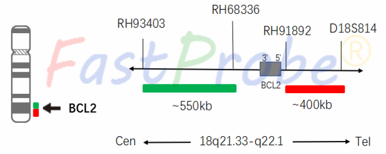
BCL2 gene break apart probe detection kit
Probe description
BCL2 is a dual-color break apart probe composed of two probes directly labeled at 18q21.33-q22.1. The green fluorescent dye labeled probe hybridizes to the proximal end of the BCL2 gene, while the orange-red fluorescent dye labeled probe hybridizes to the distal end of the BCL2 gene.
Clinical significance
Follicular lymphoma (FL) is a less malignant B cell tumor derived from the center of follicle development. FL is a common type of non-Hodgkin's lymphoma (NHL), accounting for about 10% of NHL in China and 25%-45% of NHL in Europe and America. BCL2 gene break apart and MTC gene break apart can be used both in the lymphoma diagnosis.

BCL6 gene break apart probe detection kit
Background introduction BCL6 gene is located at the 3q27 region, and the protein encoded by the BCL6 gene is a transcriptional repressor involved in the development and function of the lymphatic system. Chromosome recombination of the BCL6 gene region is present in different types of non-Hodgkin's lymphoma (NHL), including diffuse large B-cell lymphoma (DLBCL) and follicular lymphoma (FL). The most common translocation t(3;14)(q27;q 32.3) of BCL6 led to fusion of the IGH-BCL6 gene. Therefore, detection of BCL6 rearrangement by fluorescence in situ hybridization may be helpful in predicting clinical outcomes in patients with NHL (non-Hodgkin's lymphoma).
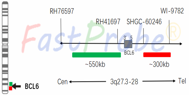
Probe description
BCL6 is a dual-color break apart probe composed of two probes directly labeled to 3q27.3-q28. The green-labeled fluorescent probe directly hybridizes with the 3q27.3 proximal BCL6 gene, while the orange-red labeled fluorescently probe directly hybridizes with the distal end of the BCL6 gene at the 3q27.3-q28 distal group.
Clinical significance
In diffuse large B-cell lymphoma, BCL6 gene can translocate with multiple genes, the incidence rate is 20%-40%; in follicular lymphoma, the incidence rate is 5-15%. Burkitt's lymphoma is morphologically suggestive of typical age, morphology, and immune characterization. If any of these three features is not typical or has a history of follicular lymphoma, and is accompanied by MYC gene breaks and BCL6 gene breaks, it should be diagnosed as a grey-area lymphoma between Burkitt and DLBCL. This probe aims to detect whether the BCL6 gene is broken and translocated. The BCL6 gene break is an independent indicator for evaluating survival rate and recovery rate.
MYC gene break apart probe detection kit
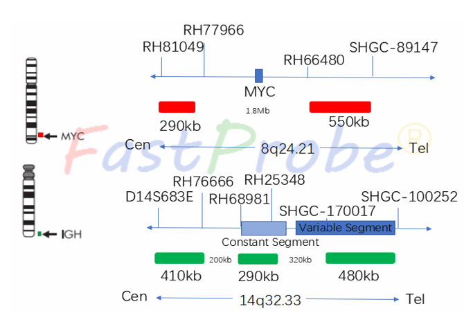
Background introduction MYC proto-oncogene is located on chromosome 8q24 and encodes a transcription factor that regulates cell growth. It is mainly activated by amplification and chromosome translocation rearrangement, and its downstream target genes affect cell proliferation, DNA and protein synthesis and metabolism. In recent years, MYC gene abnormalities have become an important indicator of poor prognosis in patients with DLBCL.
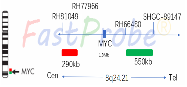
Probe description
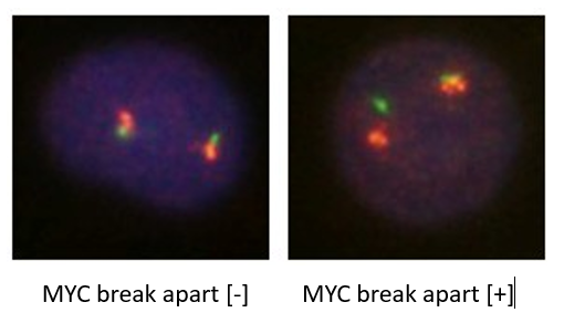
MYC dual color break apart probe is a two directly labeled hybrid probes that hybridize at the 8q24.21 region. The probe directly labeled with the orange-red fluorescent dye hybridizes with the proximal end of the MYC gene, and the green fluorescent-labeled probe hybridizes with the distal end of the MYC gene.
Clinical significance
MYC gene breaks in 5%-10% of patients with diffuse large B-cell lymphoma, and the survival time is significantly shorter than that of normal patients is. MYC gene break, BCL2 gene break or BCL6 gene break can be used for the diagnosis of double-hit lymphoma (DHL).
IGH gene break apart probe detection kit
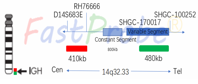
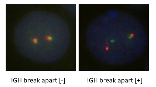
Background introduction IGH separated dual-color probe is designed to detect translocation of the IGH gene at chromosome 14q32.33. IGH gene rearrangement can be used as a specific molecular marker for detecting minimal residual disease of DLBCL. IGH gene breaks and translocations occur in 50% of B cell NHL and various other lymphomas, and can translocate with more than 50 genes.
Probe description
IGH is a dual-color break apart probe, consisting of two probes directly labeled at 14q32.33. The probe labeled with orange-red fluorescence hybridizes at the IGH gene proximal end, while the probe labeled with green fluorescence hybridizes with the distal end of the IGH gene.
Clinical significance
The fusion of the IGH gene with a variety of genes can be used for diagnosis, especially for B-cell and T-cell NHL, non-classical HL, and reactive hyperplasia that are not characterized by histopathology and immunohistochemistry. These tests are helpful for the diseases diagnosis.
MYC /IGH gene fusion probe detection kit
Background introduction MYC proto-oncogene is located on chromosome 8q24, and its encoded transcription factors are closely related to cell growth and proliferation, as well as tumorigenesis. Translocation of the MYC gene is considered to be a cytogenetic marker of Burkitt's lymphoma (BL), but is also present in other types of lymphoma. About 80% of BL cases have a translocation between the c-MYC gene locus and the Ig gene locus (t(8;14) (q24;q32)), ie, the high activity of the c-MYC translocation to the Ig locus, thus constituting a highly active genes rearrangement, initiating c-MYC transcription, enhancing c-MYC expression, promoting malignant transformation, and ultimately leading to tumorigenesis. The t(8;14) (q24;q32) test helps to diagnose Burkitt's lymphoma and can guide the treatment of high-grade B-cell lymphoma.
Probe description
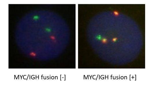
MYC /IGH is a dual-color, double-fusion probe consisting of a green fluorescent directly labeling IGH probe across known IGH breakpoint, and an orange-red fluorescence directly labeling MYC probe across known MYC breakpoint.
Clinical significance
T(8;14) can be used to assist in the diagnosis of Burkitt's lymphoma - BL- (75% incidence) and guide the treatment of high-grade B lymphoma the prognosis is poor.

BCL2/ IGH gene fusion probe detection kit
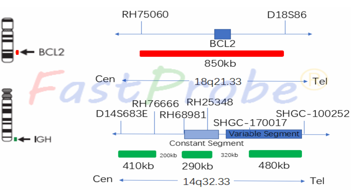
Background introduction BCL2/IGH dual-color double fusion probe is designed to detect the translocation of t(14;18)(q32.3;q21.3), that is, the IGH gene at chromosome 14q32.33 and the BCL2 gene at 18q21.33 region. The translocation of the IGH (immune sphere gene) and BCL2 (B cell lymphoma) gene involved is a cytogenetic marker of FL (follicular lymphoma). FL is one of the most common NHL (non-Hodgkin's lymphoma). The t(14;18)(q32.3;q21.3) translocation is present in approximately 80% of patients with follicular lymphoma, but it is also found in 20% to 30% of diffuse large B-cell lymphoma (DLBCL) patients. If histology is uncertain, fluorescence in situ hybridization (FISH) can be used to detect t(14;18).
Probe description
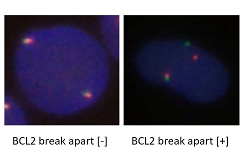
BCL2 orange probe labeled with an orange-red fluorescent dye and IGH green probe labeled with a green fluorescent dye bound to the target detection site by in situ hybridization. Under normal conditions (BCL2/IGH gene is not fused), it shows two orange-red signals and two green signals under a fluorescence microscope. When there is gene fusion, the green and orange-red signals form a yellow fusion signal due to recombination.
Clinical significance
The t(14;18) translocation occurs in 85% of follicular lymphoma (FL) and 1/3 of diffuse lymphoma (DL) with a poor prognosis. Studies have shown that BCL2/IGH translocation rearrangement plays a role in stimulating B lymphocyte hyper proliferation. The incidence of most translocations in patients with non-Hodgkin's lymphoma is significantly higher than that in healthy controls
CCND1 (BCL1)/IGH gene fusion probe detection kit
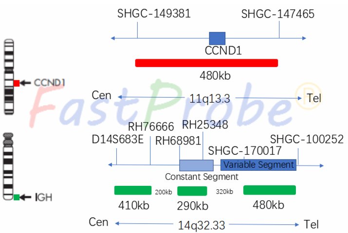
Background introduction CCND1/IGH dual-color fusion probe is used to detect t(11;14) (q13.3; q32.3) translocations in up to 95% of mantle cell lymphomas (MCLs). At the same time, t(11;14) is also present in other lymphoproliferative diseases, such as juvenile lymphoblastic leukemia (PLL) and plasma cell myeloma.
Probe description
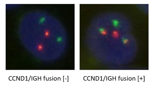
CCND1 probe labeled with an orange-red fluorescent dye and IGH green probe labeled with a green fluorescent dye bound to the target detection site by in situ hybridization. Under normal conditions (CCND1/IGH gene is not fused), it shows two orange-red signals and two green signals under a fluorescence microscope. When there is a gene fusion, the green and orange signals recombine to form yellow fusion signal.
Clinical significance
Mantle cell lymphoma is a subtype of NHL with poor prognosis; t(11;14)(q13.3;q32.3) can be used for the auxiliary diagnosis of mantle cell lymphoma (MCL). It can also be used for the MCL and CLL differentiation.
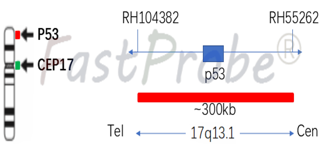
P53 gene probe detection kit
Background introduction P53 gene is highly correlated with human tumors and is an important gene tumor suppressor. The 53kD protein encoded by the P53 gene plays an important regulatory role in the cell cycle, has a growth inhibitory effect under normal conditions, and plays an important role in DNA cell damage response, cell death and differentiation in the cell cycle.
Probe description
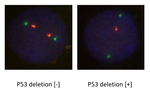
P53 gene probe uses an orange-red dye to label P53 gene region, and a green dye to label chromosome 17-centromere region (CEP17). P53 gene marker region is located at 17q13.1, and CEP17 probe is labeled with a specific alpha satellite sequence.
Clinical significance
P53 gene deletion indicates patient’s poor response to chemo-radiotherapy and are prone to metastasis, which can be used as an indicator for therapeutic efficacy and prognosis. If p53 gene mutation occurs in the early stage of tumorigenesis, it will be helpful for the tumor early diagnosis.