IGH gene (encoding the immunoglobulin heavy chain) rearrangement has been proved to be an early event in the MM ladder molecular pathogenesis, usually occurring at the 14q32 region. The breakpoints are mainly in the D and J regions, occurring in about 50% to 60% of MM patients. Partner chromosomes of the IGH gene translocation mainly include 11q13 (BCL1/CCND1), 4p16.3 (FGFR3), 16q23 (MAF), 20q11 (MAFB) and 6p21 (CCND3).
Dwarka Sector-7, New Delhi-110075
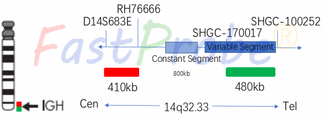

IGH gene break apart detection probe kit
Probe description
IGH is a dual-color break apart probe consisting of two probes directly labeled at 14q32.33, in which the orange-red fluorescent-labeled probe hybridizes to the proximal end of the IGH gene, while the green fluorescent-labeled probe hybridizes to the IGH gene distal end.
Clinical significance
IGH gene break and translocation types are complex and involve multiple genes, commonly found in ALL/MM/lymphoma; can be used to detect abnormalities and minimal residual lesions of IGH gene; IGH gene break apart can be used as a marker for malignant cloning of myeloma cells, not affected by clinical stages and immune types, it can be used as a strong basis for MM diagnosis.
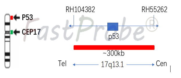
P53 gene detection probe kit
Background introduction P53 gene is highly correlated with human tumors and is an important gene tumor suppressor. The 53kD protein encoded by the P53 gene plays an important regulatory role in the cell cycle, and has a growth inhibitory effect under normal conditions, and plays an important role in DNA cell damage response, cell death and differentiation in the cell cycle.
Probe description
P53 gene probe uses an orange-red dye to label P53 gene region, and a green dye to label chromosome 17-centromere region (CEP17). P53 gene marker region is located at 17q13.1, and CEP17 probe is labeled with a specific alpha satellite sequence.
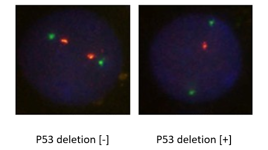
Clinical significance
P53 deletion occurs in 1/3 of newly diagnosed MM (Multiple myeloma), which means short survival and poor prognosis for patients receiving conventional chemotherapy dose.
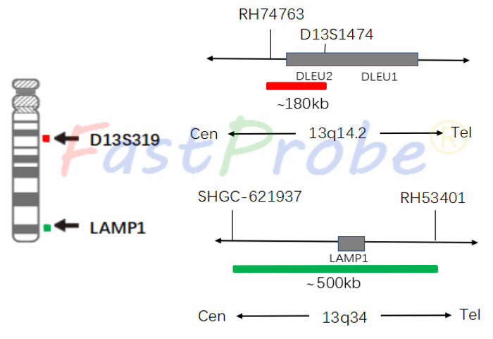
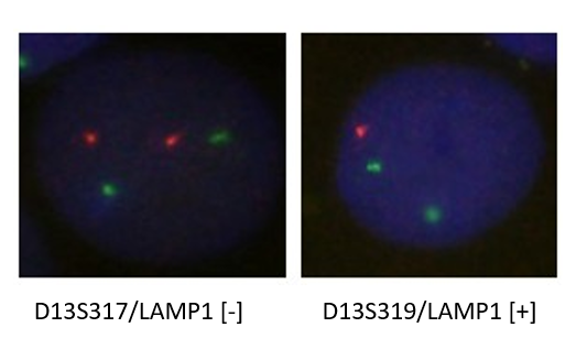
13q14/13q34 (D13S319/LAMP1) gene deletion detection probe kit
Background introduction Multiple myeloma (MM) is one of the most common malignant plasma cell diseases, accounting for 10% of hematopoietic malignancies. It is characterized by malignant proliferation of monoclonal plasma cells and secretion of a large number of monoclonal immunoglobulins, causing a series of clinical changes such as bone pain, pathological fracture, hematopoietic abnormalities, monoclonal globulinemia and impaired renal function. The current general MM diagnostic criteria are mainly standard WHO (2001) and International MM Working Group (2003). This disease is easily misdiagnosed and the rate of misdiagnosis is as high as 60%. Clinical studies have found that the development of MM is accompanied by changes in the number or structure of related genes at various specific cytogenetic levels. For example, chromosome 13 occurs in 85% of MM patients.
Probe description
13q14/13q34 is a dual-color hybrid probe. The orange-red fluorescent dye directly labels the D13S319 probe and specifically detects the D13S319 gene at 13q14.2. The green fluorescent dye directly labels the 13q34 probe, which specifically detects LAMP1 gene at the 13q34 region.
Clinical significance
Clinical studies have found that the occurrence and development of MM is accompanied by a variety of specific changes in the number or structure of related genes at the cytogenetic level. Chromosome 13 haplotypes occur in 85% of patients with MM, and adverse prognostic factor found.
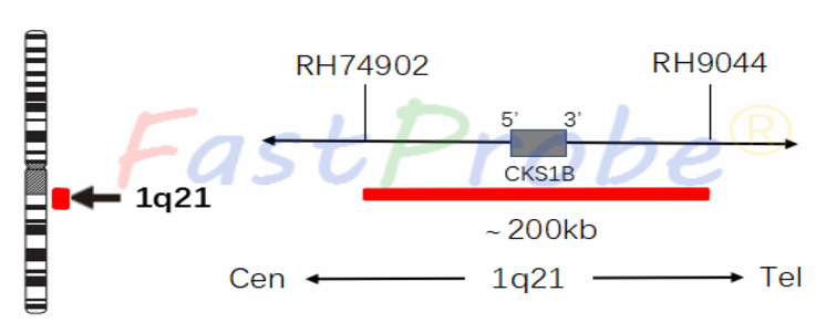
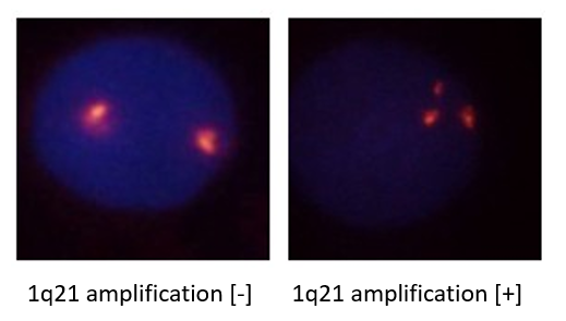
1q21 gene amplification detection probe kit
Background introduction Chromosome 1 abnormality is one of the most common cytogenetic findings in MM (Multiple myeloma). A major feature of B cell malignancies is the slow increase in malignant plasma cells grown in the bone marrow. The CKS1B gene is located at 1q21 of chromosome 1 long arm end. In the progression of myeloma disease, tandem repetition and skip translocations of the 1q21 band occur, whereas in patients with multiple myeloma, 1q amplification is associated with poor prognosis.
Probe description
1q21 gene amplification detection probe uses an orange-red fluorescent label 1q21 region, and the 1q21 probe binds to the target detection site by in situ hybridization. This method is used to detect abnormalities of multiple myeloma genes, and provide clinical reference for the differentiation, prognosis and medication for leukemia patients.
Clinical significance
1q21 (CKS1B) is the most common genetic abnormality in MM. The expansion of CKS1B gene leads to the up-regulation of cell cycle, which causes many proliferative diseases. 1q21 amplification is often associated with MM phenotype infiltration, poor prognosis and rapid disease progress.
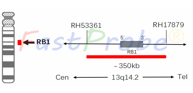
RB1 deletion detection probe kit
Background introduction RB1 gene is located in the 13q14.2 region, its encoded protein acts as a tumor suppressor and plays a very important role in cell cycle and genomic DNA stability.
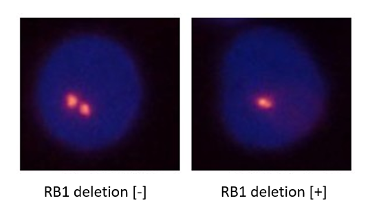
Probe description
RB1 gene deletion detection probe uses an orange-red fluorescent label RB1 gene, and the RB1 probe bind to the target detection site by in situ hybridization. This method is used to detect the abnormalities in multiple myeloma genes, and provide clinical reference for the differentiation, prognosis and medication for leukemia patients.
Clinical significance
Some reseachers recommend MM differential diagnosis at the cytogenetic level. These changes are closely related to the prognosis of patients. Patients with RB1 gene deletion have a moderate prognosis with a median survival of 40 months.
CCND1/IGH gene fusion detection probe kit
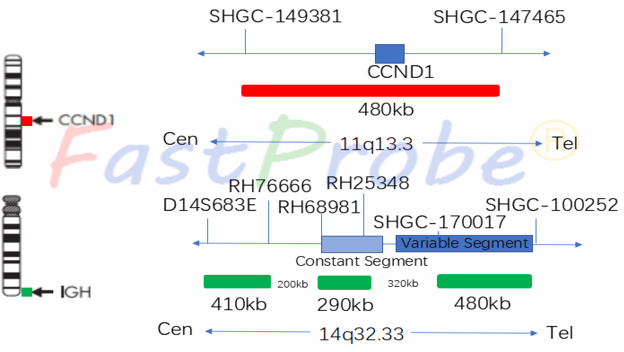
Background introduction CCND1/IGH dual-color double fusion probe is used to detect the translocation of t(11;14)(q13.3;q32.3) which often occurs in MM. This translocation exists in the CCND1 gene near the IGH (immunoglobulin heavy chain) gene, which leads to overexpression of the CCDN1 gene. Detection of t (11; 14) translocation has important clinical significance.
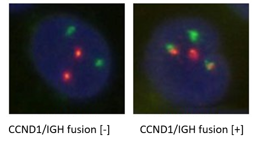
Probe description
CCND1/IGH is a dual-color, double-fusion probe with an orange-red fluorescent dye directly labeled with the CCND1 probe and a green fluorescent directly labeled IGH probe. Under normal conditions (CCND1/IGH gene did not fuse), it shows two orange-red signals and two green signals under a fluorescence microscope. When there is gene fusion, the green and orange-red signals form a yellow fusion signal as recombination result.
Clinical significance
t(11;14) is one of the most common abnormal translocations in MM. MM patients with t(11;14) translocation or no other genetic changes have a good prognosis, with a median survival of 50 months.