Background introduction Human epidermal growth factor receptor 2 (HER2, also known as ERBB2, Neu, ErbB-2, CD340 or p185) is a proto-oncogene HER2/neu located on the long arm 17q12 of human chromosome 17. The coding, which is a member of the epidermal growth factor receptor (EGFR/ErbB) family, has tyrosine kinase activity and is involved in signal transduction of cell growth and differentiation. The oncogenic mechanism of the HER2 oncogene includes inhibition of apoptosis, promotion of cell proliferation, increase of invasiveness of tumor cells, and promotion of tumor vascular and lymphangiogenesis. 20% of breast cancer and 12% of gastric cancer patients showed positive HER2 gene amplification.
Dwarka Sector-7, New Delhi-110075
HER2 gene amplification detection probe kit.
Probe description
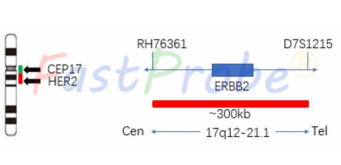
The HER2 gene amplification probe uses the orange-red dye to label the HER2 gene region, and the green dye is used to label the chromosome 17 centromere region (CEP17). The HER2 gene marker region is located at 17q12-q21.1, and the CEP17 probe adopts an alpha satellite sequence, which has extremely high specificity and does not hybridize with other chromosome centromeres to produce noisy spots.
Clinical significance
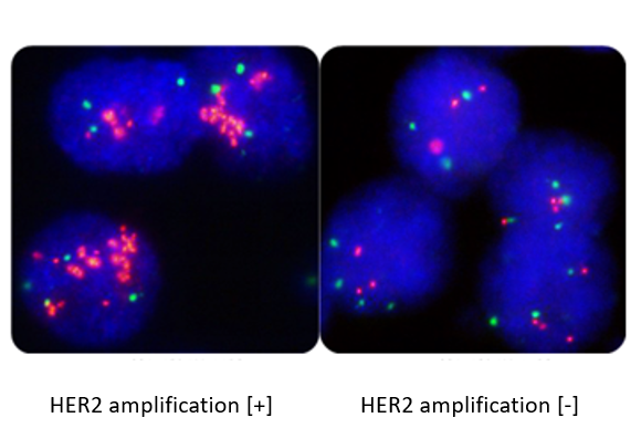
Fluorescence in situ hybridization (FISH) is a clinically recognized “gold standard” for HER2 detection. It can accurately and repeatedly evaluate the status of HER2 gene in cancer cells. Compared with IHC, FISH has higher consistency. The patients with positive HER2 gene amplification were effectively treated with targeted drugs such as monoclonal antibodies Herceptin and Lapatinib. The prognosis of patients with positive HER2 gene amplification was poor, and the disease-free survival and overall survival were significantly shortened.
TOP2A gene amplification detection probe kit
Background introduction TOP2A gene encodes a DNA topoisomerase that participates in processes such as chromosomal concentration, chromatid separation, and release of torsional stress during DNA transcription and replication. The gene encoding this form, TOP2α, is located on chromosome 17, the beta gene located on chromosome 3, and multiple mutations in the TOP2α gene are involved in development.
Probe description
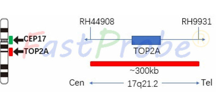
TOP2A gene amplification probe uses the orange-red dye to label the TOP2A gene region, and the green dye to label the chromosome 17 centromere region (CEP17). TOP2A gene-labeled region is located at 17q21.2, and the CEP17 probe adopts an alpha satellite sequence, which has extremely high specificity and does not hybridize with other chromosome centromeres to produce noisy spots.
Clinical significance
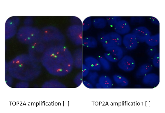
Patients with abnormal TOP2A gene indicates a shorter recurrence-free survival, and patients with TOP2A gene deletion have a worse prognosis. In the study of advanced breast cancer, it was found that the abnormality of TOP2A gene was significantly correlated with the protein expression and the sensitivity of tumor cells to anthracyclines. Therefore, the detection of TOP2A gene status has guiding significance for the treatment and prognosis of breast cancer.
MYC gene amplification detection probe kit
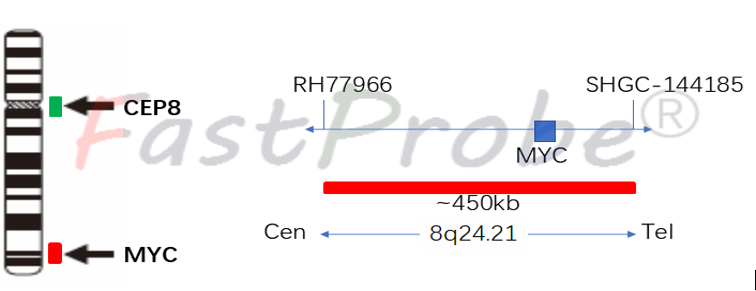
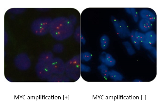
Background introduction The MYC proto-oncogene is located on chromosome 8q24 and encodes a transcription factor that regulates cell growth. It is activated mainly by amplification and chromosome translocation rearrangement. MYC gene amplification is associated with the development of a variety of tumors (including breast cancer, colon cancer, lung cancer, hematopoietic tumors, etc.).
Probe description
MYC gene amplification probe uses orange-red dye to label MYC gene region, and green dye to label chromosome 8 centromere region (CEP8). The MYC gene marker region is located at 8q24.21, and the CEP8 probe is labeled with a specific alpha satellite sequence.
Clinical significance
MYC gene amplification is a common phenomenon in tumors and can be found in a variety of malignant tumors such as breast cancer, nasopharyngeal cancer, and cervical cancer. The prognosis of breast cancer patients with MYC gene amplification is poor.