Background introduction Bladder cancer is the most common malignant tumor of the urinary system. It is more common in men and the incidence is about 4 times that of women. The average age of onset is 65 years. Seventy-five percent of the new cases are superficial tumors, of which 50-80% will have recurrences one to many times after treatment; 15-25% will progress to invasive cancer. Therefore, patients with superficial bladder cancer need to pay close attention to the recurrence and deterioration of the tumor. Cystoscopy or urine exfoliative cytology is recommended for patients with hematuria over 40 years of age. Probe description
Dwarka Sector-7, New Delhi-110075
Bladder cancer probe detection kit
However, cystoscopy can cause unnecessary pain to the patient
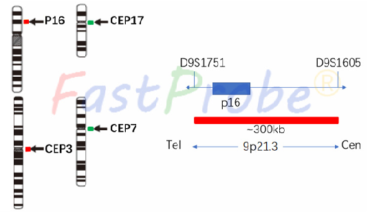
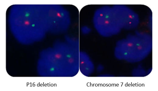
However, cystoscopy can cause unnecessary pain to the patient, and because of the stimulation of the bladder wall tumor, it will cause the malignant expansion and metastasis of the tumor, which is not suitable for large-scale screening, and the cytological examination is insufficiently sensitive. Fluorescence in situ hybridization detection of urine sediment cells showed strong advantages in early diagnosis and postoperative recurrence of bladder cancer. Bladder cancer probes consist of two groups of probes. The orange dye is used to label the P16 gene region, the green dye is used to label the centromere region of chromosome 17 (CEP17); the orange dye is used to label the centromere region of chromosome 3 (CEP3), and the green dye is used to label the centromere region of chromosome 7 (CEP7). The P16 gene marker region is located at 9p21.3, and the chromosomal centromere probes are labeled with a specific alpha satellite sequences. Clinical significance The most common genetic alteration of urinary transitional epithelial cell carcinoma is the partial or total loss of chromosome 9 (e.g. p16 locus). In addition, the development of urinary transitional epithelial cell carcinoma is closely linked to chromosomal instability. In particular, it is closely related to the aneuploidy of chromosomes 3, 7, and 17. FISH is a non-invasive test, which can detect exfoliated cells in patient’s urine. If there are two or more abnormalities in the above four indicators, or if one of the indicators has a complex abnormality, it can be determined as the urinary system transitional epithelial cell carcinoma.
P53 gene probe detection kit
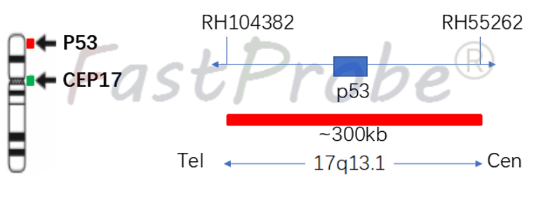
Background introduction The P53 gene is highly correlated with human tumors and is an important tumor suppressor gene. The 53kD protein encoded by the P53 gene plays an important regulatory role in the cell cycle, and has a growth inhibitory effect under normal conditions, and plays an important role in DNA cell damage response, cell death and differentiation in the cell cycle. Probe description P53 gene amplification probe uses an orange-red dye to label the P53 gene region, and a green dye to label chromosome 17 centromere region (CEP17). P53 gene marker region is located at 17q13.1, and the CEP17 probe is labeled with a specific alpha satellite sequence.
Clinical significance
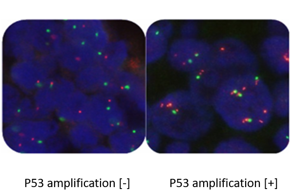
P53 gene amplification and deletion indicate tumor poor prognosis, its insensitivity to conventional chemotherapy, and is inclined to metastasis.