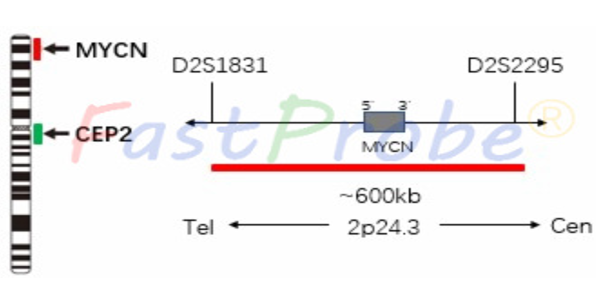
Background introduction MYCN gene is located in the p24.3 region of chromosome 2 and encodes a 62-64 kDa transcription factor. MYCN is mainly expressed in the nervous system. Probe description MYCN gene amplification probe uses an orange-red dye to mark the MYCN gene region, and a green dye to label the chromosome 2 centromere region (CEP2). The MYCN gene marker region is located at 2p24.3, and the CEP2 probe is labeled with a specific alpha satellite sequence.
Dwarka Sector-7, New Delhi-110075
MYC gene amplification detection probe kit
Clinical significance
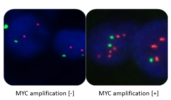
MYCN gene amplification occurs in approximately 25% of patients with neuroblastoma. MYCN gene amplification is associated with infiltration, metastasis and poor prognosis of neuroblastoma. When the MYCN gene amplification factor is less than 10, the clinical treatment plan may not be treated after the complete removal of the primary tumor; when the MYCN gene amplification factor is >10, the conventional chemotherapy should be performed for 12 months after the surgical resection, and local radiotherapy is needed if necessary.
MLL (KMT2A) gene detection probe kit
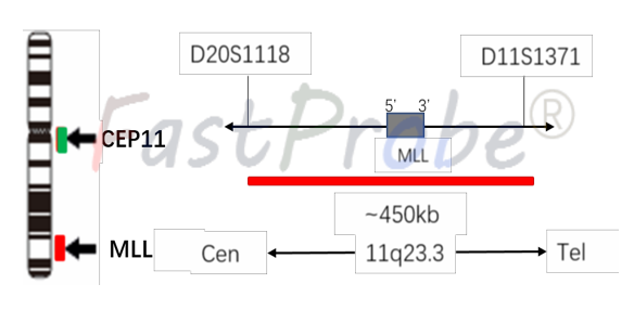
Background introduction The MLL (KMT2A) gene is located in the q23.3 region of chromosome 11, which encodes a transcriptional coactivator that plays an important role in the regulation of gene expression during early development and hematopoiesis. Probe description The MLL (KMT2A) gene detection probe uses an orange-red dye to label the MLL gene, and a green dye to label chromosome 11 centromere region (CEP11). The MLL (KMT2A) gene marker region is located at 11q23.3, and the CEP11 probe is labeled with a specific alpha satellite sequence.
Clinical significance
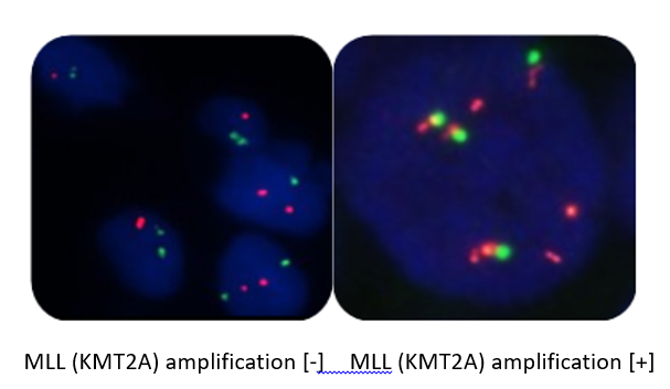
MLL (KMT2A) gene deletion is seen in primary neuroblastoma, and MLL (KMT2A) inactivation is associated with malignant progression of neuroblastoma in malignant progression of neuroblastoma without MYCN gene amplification.
MDM4 amplification detection probe kit
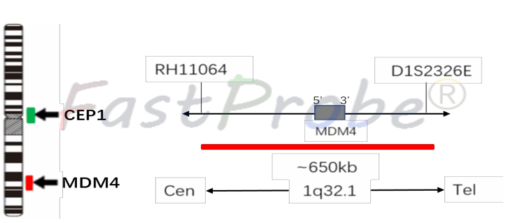
Background introduction MDM4 (HDMX, MDMX) gene is located in the q32.1 region of chromosome 1, encoding a protein containing 490 amino acid residues. MDM4 is an important regulator of p53 upstream and plays a major role in apoptosis. Probe description MDM4 (HDMX, MDMX) gene amplification probe uses an orange-red dye to label MDM4 gene region, and a green dye to label the chromosome 1 centromere region (CEP1). MDM4 (HDMX, MDMX) gene marker region is located at 1q32.1, and the CEP1 probe is labeled with a specific alpha satellite sequence.
Clinical significance
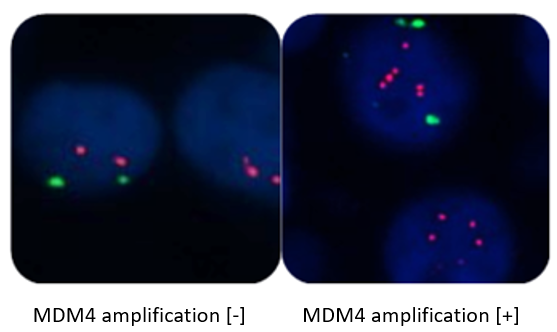
MDM4 amplification is seen in 65% of primary neuroblastomas, MDM4 is a primary neuroblastoma-specific chemotherapy target, and MDM4 gene amplification patients are not sensitive to chemotherapy.
1p36 (SRD) gene deletion detection probe kit
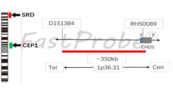
Background introduction Deletion of the 1p36 region (SRD gene) can occur in a variety of tumors, such as neuroblastoma, glioma, leukemia, lymphoma, and the like. Probe description 1p36 (SRD) gene deletion probe uses an orange-red dye to label the SRD gene region, and a green dye to label chromosome 1 centromere region (CEP1). SRD gene marker region is located at 1p36, and the CEP1 probe is labeled with a specific alpha satellite sequence.
Clinical significance
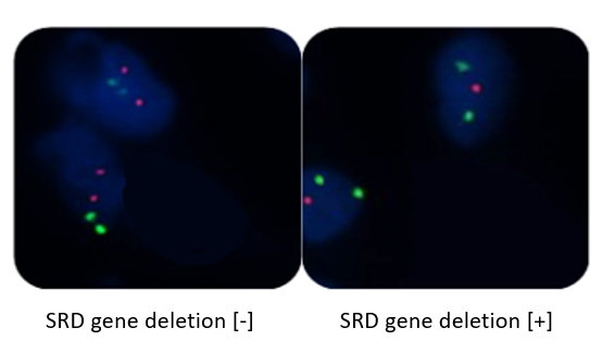
The deletion of 1p36 (SRD gene) in neuroblastoma is the most typical genetic alteration. The detection of 1p36 heterozygous deletion has a major significance in the clinical guidance and prognosis of neuroblastoma. 1p36 patients with neuroblastoma are prone to recurrence, have a poor prognosis, and are sensitive to chemotherapy.